

 |
0/0 |


- Copyright : CDC / IMAGE POINT FR / BSIP
- Collection : imagepointfr
- Model release : Sans objet
- Property release : Sans objet
- Autorisations : Toute utilisation
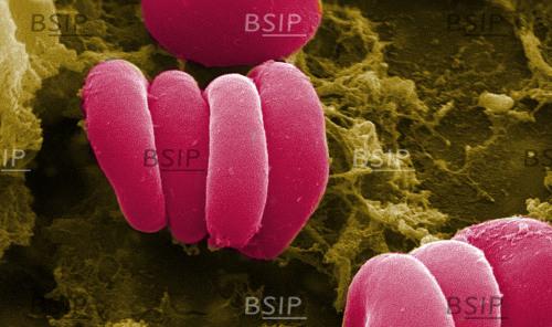
Légende : This scanning electron micrograph (SEM) depicted a closer view of a number of red blood cells found enmeshed in a fibrinous matrix on the luminal surface of an indwelling vascular; Magnified 7766x. In this instance, the indwelling catheter was a tube that was left in place creating a patent portal directly into a blood vessel. Some of the erythrocytes are grouped in a stack known as a "Rouleaux formation". Note the biconcave cytomorphologic shape of each erythrocyte, which increases the surface area of these hemoglobin-filled cells, thereby, promoting a greater degree of gas exchange, which is their primary function in an in vivo setting. In their adult phase, these cells possess no nucleus. What appears to be irregularly-shaped chunks of debris, are actually fibrin clumps, which when inside the living organism, functions as a key component in the process of blood clot formation, acting to entrap the red blood cells in a mesh-like latticework of proteinaceous strands, thereby, stabilizing and strengthening the clot, in much the same way as rebar acts to strengthen, and reinforce cement.